
Seminar Transcript | Legal Medical Diagnosis Requirements in the UK: 2019 Diagnosis, Education, and Rehabilitation of Cerebral Visual Impairment
Last edited: 2026-07-27
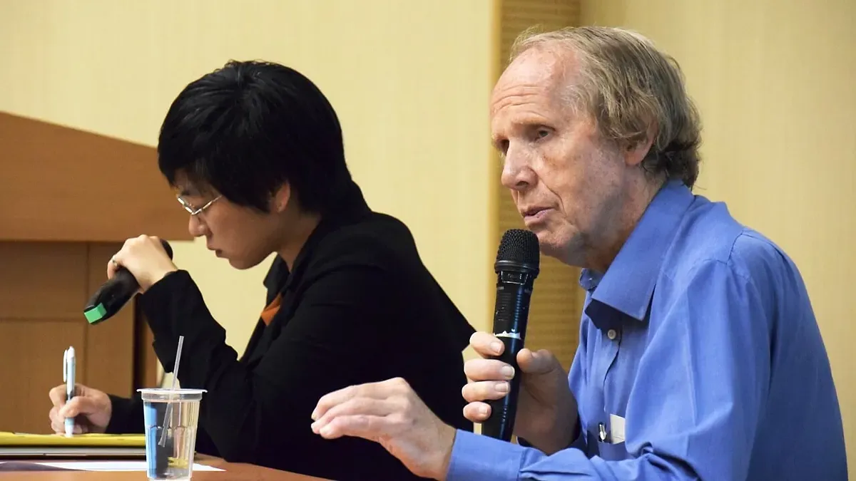
Editor's Note: This article is compiled from the contents of the "Seminar on the Diagnosis, Education, and Rehabilitation of Cerebral Visual Impairment" held on April 27.
The keynote speaker, Gordon N. Dutton, MD, is a Doctor of Medicine, a Fellow of the Royal College of Ophthalmologists (UK), and an Honorary Fellow of the Royal College of Surgeons of Edinburgh. He served as a pediatric ophthalmologist at the Royal Hospital for Sick Children in Glasgow, Scotland for over 30 years, and currently serves as an honorary professor in the Department of Vision Sciences at Glasgow Caledonian University and an honorary research fellow at the University of Glasgow. He is widely recognized for his work on childhood visual impairments caused by brain injury, and has long served as a committee member and consultant for organizations across the UK, North America, and elsewhere, including as a visiting professor at Boston Children's Hospital.
The topic I am reporting on today is "Legal Medical Diagnosis Requirements in the UK." While I may not be entirely familiar with this specific subject, I will discuss what we can do within this context from the perspective of a physician.
"Diagnosis for the Patient" vs. "Diagnosis for Those Who 'Do Not Believe'"
When we talk about diagnosis, diagnosing a witness versus providing a diagnosis for those who "do not believe the matter" are two different things. In clinical practice in the UK, its purpose is to help people. Scientific practice has a very different purpose from clinical practice, because scientific practice aims to provide information, educate, and conduct detailed validation of discovered findings.
- Clinical practice aims to help people
臨床實踐旨在幫助人們 - Scientific practice aims to inform and teach, and needs detailed validation of findings
科學實踐旨在提供資訊和教學,並需要對結果進行詳細驗證 - Legal practice aims to have one of two forms of proof
法律實踐旨在上述的兩種證明形式中取捨- Civil cases – Balance of probabilities (50:50)
民事案件 - 均衡的可能性(50:50) - Criminal cases – Beyond reasonable doubt (99:1)
刑事案件 - 超越合理懷疑(99比1)
- Civil cases – Balance of probabilities (50:50)
As a doctor, in legal practice I also encounter two different forms of court cases or have to provide two different forms of evidence. In civil cases, the focus is on finding a balance; the burden of proof in civil matters is slightly lower, at a 50:50 ratio. However, in criminal cases, much stronger evidence is required to counter the arguments presented by the opposing side, so the standard here is much higher, akin to a 99:1 ratio. When discussing diagnosis in a clinical setting, the starting point is that the witness believes in the medical expertise you possess. In science, however, much like all scientific principles, the initial starting point is "I do not believe you, so I must find evidence to verify it."
When it comes to the law, we enter a different dimension, especially in adversarial courts where two opposing camps are present from the outset: the prosecution, which may disbelieve the evidence you present, and the defense, which believes it.
- Clinical practice starts with… belief in medical skills.
臨床實踐始於......對醫學技術的信任 - Scientific practice starts with… I don’t believe you.
科學實踐始於......我不相信你 - Adversarial legal practice starts with… two camps:Disbelief by the ‘Prosecution’ and belief by the ‘Defence’
對抗性法律實踐始於...... 雙方陣營: 「對"檢方"的懷疑」 與 「對"辯方"的信任」
I know that the systems in the UK and France differ somewhat. The UK judicial system adopts an adversarial approach in court, whereas France uses an inquisitorial approach, meaning its goal is to weigh different information to determine what the truth actually is. However, in a place like the UK which adopts an adversarial system, the defense must overcome the prosecution's disbelief regarding the evidence presented by the defense. This actually stems from the dialectical communication methods of ancient Greece: the prosecution presents its arguments regarding the defendant's guilt, and the defense must then present counter-arguments explaining why the defendant is innocent. After hearing both the affirmative and negative arguments, the judge or jury reaches a conclusion—a process of thesis, antithesis, and conclusion. The judge or jury relies on two forms of evidence to render a verdict.
Yet, a verdict is not necessarily "the truth." Being pronounced guilty or not guilty does not always align with actually being guilty or innocent. Guilt and innocence carry two different meanings; for example, one type of guilt is being "truly guilty," while another is being "found guilty in a court of law." Thus, these are two different types of guilt. The same applies to innocence: one is being truly innocent, and the other is the court finding you innocent.
In the UK justice system, there have also been miscarriages of justice where certain evidence was misinterpreted or presented incorrectly, resulting in innocent individuals being convicted. This is why, from my perspective, there should be no death penalty, because if an individual is actually innocent yet the state imposes such a punishment, the state has committed murder. I must also emphasize that this viewpoint is strictly my own as a physician; it is not a legal viewpoint.
The UK - Adversarial Courts
As a physician, I have actually served as a witness for both opposing camps in court—for instance, testifying to prove the innocence of the accused or to prove guilt, meaning I have appeared in court on behalf of either the prosecution or the defense. In an adversarial court system like that of the UK, evidence comes in two forms—though I actually believe there are three, which I will explain shortly.
Medical evidence comes in two forms…
醫學證據有兩種形式......
- The professional witness: provides evidence based on the facts within the case
專家證人(The professional witness):根據案件中的事實提供證據 - The expert witness: provides evidence based on the following
專業證人(The expert witness):根據以下提供證據
- Interpretation of the case
為案件找出解答 - Expert opinion (has to prove expertise)
專家意見 (必須證明具備專業知識)
- Interpretation of the case
The most common form of court evidence is provided by the professional witness. In such a scenario, a doctor provides evidence based strictly on the facts. The other is the expert witness. An expert witness does not testify in the capacity of the patient's treating physician; this expert witness may be a doctor or an engineer, depending on the type of evidence required in the case. Upon taking the stand, the expert witness must present expert information while also considering past literature and specialized knowledge in the field. An expert witness offers an interpretation of the case and provides expert opinion, but must also prove that they indeed possess expertise in that specific field. Recently in the UK, we have also seen instances where so-called expert witnesses became entangled in litigation themselves because these individuals were not genuine experts.
Based on my experience, physicians are also frequently manipulated, and I have personally experienced this twice. On one occasion, I took the stand as a professional witness. I had flown back to Scotland from northern India to testify, and the subject of the testimony was my patient, meaning I was acting as a professional witness providing testimony. The case involved non-accidental injury to a toddler. The prosecution in the Scottish court insisted on having me testify as an expert witness. The prosecutor asked me questions treating me as an expert witness, so I turned to the judge and said, "Your Honor, I am not an expert witness on this subject today; I am a professional witness, and therefore I refuse to answer the question just asked." The judge accepted my stance, and the flustered prosecutor stated he had no further questions for the witness, whereupon I flew back to northern India. The prosecutor in this case was clearly abusing his power.
The third distinct form of evidence occurred when I was asked to testify on a medical and pharmaceutical subject. When asked a related question, I turned to the judge and said, "In answering this question, I am acting neither as a professional witness nor as an expert witness, but I can answer it because I studied this in university, so I know the answer. If you are willing for me to adopt this stance to educate the court on this knowledge, I am willing to share this information with you and the court like a teacher, but this stance does not stem from the statements of a professional witness or expert witness." The judge accepted this approach, and I proceeded to explain the subject to the court.
Purposes of Diagnosis for Patients
- Determine the cause of the condition (anatomical and/or pathological)
確定病因(解剖 和/或 病理) - Provide corresponding treatment or condition management
提供相應的治療或病情控制
Let us return to the subject of diagnosis. A diagnosis made "for the patient" and a diagnosis made "for those who do not believe" are vastly different. First, let us look at diagnosis made for the patient: its purpose is to determine the cause of these symptoms. We may identify why these symptoms are present from an anatomical or pathological perspective. Another purpose is to provide appropriate treatment or manage the patient's condition. This is a diagnosis built on the patient's trust, and the physician also believes it to be a correct diagnosis. As ophthalmologists, we can arguably examine patients faster and see more patients than any other department in the UK—we can complete an examination in roughly 5 to 10 minutes. Our diagnoses are based on our knowledge, observation, and listening to the patient's explanation, without running numerous tests.
Purposes of Diagnosis for "Research" and "Courts"
- Identify the causes of "symptoms" and "signs"
找出「症狀」和「體徵」的原因 - Provide explanations, e.g., providing explanations for behavior
提供解釋,例如:為行為提供解釋 →Entails a higher burden of proof
→具有更高的舉證責任
When making a diagnosis for research purposes or for a court, the objective is to identify the underlying causes of the symptoms and signs, and then provide corresponding explanations—for instance, explaining certain behaviors or symptoms. However, the difference here is that the burden of proof is much higher. Because we are not merely diagnosing a patient—where the patient deeply trusts you—diagnosing for research or the court entails a much higher burden of proof, requiring you to present much more robust evidence. From a professional standpoint, my own diagnoses in such contexts differ greatly from clinical scenarios.
The Case of Milena
Let me introduce a very important case. This patient's name is Milena. Over 20 years ago, around 32 years of age, Milena was an ophthalmic secretary who suffered from a severe respiratory illness and subsequently fell into a coma with extremely low blood pressure. She was admitted to the intensive care unit and woke up after being in a coma for 27 days. However, she experienced brain swelling. Imagine a garden watering hose: two blood vessels at the back of her brain were like a watering hose that had been sharply folded over, resulting in blindness due to ischemia in the posterior occipital lobes of her brain. As mentioned earlier, she was an ophthalmic secretary, and she was examined at our clinic two years after the incident.
I saw her in 1998 and learned that between the onset of her illness and when I met her, she had the following medical history: one year after her injury, she began to see moving objects, though for the entire year prior to that, she was completely blind and unable to see anything. This ability to be "blind yet see movement" is a phenomenon we call the "Riddoch Phenomenon."
"Riddoch" comes from the name of a British physician who observed soldiers during wartime who had suffered trauma to the rear of the brain, such as being struck by bullets in the posterior brain. These soldiers were able to see moving objects, just like this patient. He wrote a paper on this in 1917, which was a landmark case, but because it was so extraordinary, people did not believe it was real. For me, however, I did not need to run any tests during my clinical diagnosis; within two minutes, through my knowledge, I was able to arrive at a diagnosis. I told the patient she was extremely fortunate, and that what she was experiencing was the aforementioned "Riddoch Phenomenon." The patient was able to perceive colors, provided that those colors were in motion. For example, if I moved a shirt, she could see that it was blue, but if I held the shirt still, she could no longer see it. When I met Ching-k'ai yesterday, his manifestations were identical—the exact same.
The aforementioned patient Milena could see raindrops falling on a window, but could not see through the window to the scenery outside. She could see flowing water, but could not see the trees in front of her; she could also see water draining in a bathtub, but could not see her daughter sitting in the tub.
 Video 1 / Seeing raindrops falling on the window, but unable to see through to the scenery outside
 Video 2 / Seeing water drain in the bathtub, but unable to see the daughter sitting in the tub
 Figure 3 / Milena's perceptible visual fields in both left and right eyes
When diagnosing her, I found that the patient lacked conscious vision, but when I moved my fingers in front of her, she could perceive them. Figure 3 illustrates the visual fields she could perceive with her left and right eyes: red represents her right eye, and blue represents her left eye. Within the marked areas, she could perceive my fingers moving. The regions perceptible by both eyes were remarkably similar. Having similar visual field patterns in both eyes suggests that the patient sustained damage to the brain.
I had read Dr. George Riddoch's 1917 paper and found it fascinating. Because I had read it, I knew that this patient could walk around objects. Therefore, I proceeded to ask the patient to walk around three chairs in my consulting room continuously in a figure-eight pattern. The patient told me she could not see the chairs. I told her that once she started walking, she would see them. The patient said she could not do it; I replied that I believed she could. The patient said she would try, so she began to walk, and she successfully navigated around them despite the limited space, and with a charming Scottish accent said, "I didn't know I could do that!"
 Figure 4 / Computed tomography (CT) scan at the time showing bilateral ischemic changes in the occipital lobes
After her visit, we performed a computed tomography (CT) scan. The lower portion of Figure 4 shows bilateral changes in the occipital lobes due to ischemia, although the CT scanning technology at the time was not as advanced as it is today. My recommendation to her following the diagnosis was also very simple: I wrote on her prescription to buy a rocking chair so she could practice how to see things, because her body could see things once it was in motion. In fact, some children with CVI are found to enjoy rocking their bodies. I believe they make this motion precisely because they discover it helps them see.
A few months later, I read this book, which I highly recommend and suggest everyone buy and read. The authors are David Milner and Melvyn A. Goodale, and the book is titled The Visual Brain in Action. This book transformed our understanding of vision and is, in my opinion, the most important book in the field of vision over the past decade. Reading this book helped me understand why this patient possessed motor awareness and visually guided action. It was based on a specific case where the authors spent hundreds of hours studying a subject whose condition was almost identical to that of my previous patient, Milena. The patient in the book suffered brain damage from poisoning, and the researchers studied how this patient was able to navigate a visual world despite having no eyesight. Conceptually, her experience was the same as Ching-k'ai's.
 Figure 5 / The Visual Brain in Action
 Figure 6 / Sight Unseen
Shortly afterward, I read another, slimmer book by the same authors that discussed similar matters in a simpler way. This book mentioned "air writing," where a patient could trace out the shapes of letters in the air that they could not see. Later, the patient used a different method—not actual writing, but imagining tracing out with their finger the letters they could not actually see. I subsequently asked Milena if she could do the same thing, and she traced the letters O and N in the air. While tracing these letters with her finger, she said she did not know how she managed to do it. I replied that I did not know how she did it either, but I later understood, and I will explain shortly. Six months later, Milena also learned how to read through imagination—that is, reading letters by imagining her finger moving. Thus, by slightly moving her head and imagining her finger tracing the shapes of the letters, she could read newspaper headlines. Therefore, even though she lacked the visual brain, she still found a way to read.
Although Milena lacked a visual brain, she was still able to catch a ball rolled toward her. In the video, Milena says, "Is this ball smaller than the previous one? But I didn't know it was smaller until I caught it." So I wrote to the two authors I just mentioned and told them, "I've read your book, but I now have a patient even better than yours; would you like to meet her?" They replied that of course they would. So when I met Milena, I asked her if she would like to take a vacation to Canada, where researchers would conduct some studies on her, followed by a week of vacation with her husband and daughter. I will play a video they filmed in Canada in a moment. The researchers in Canada conducted extremely in-depth research on Milena; let us watch the video they made.
Video / The Blind Woman Who Saw Rain | SKUNK BEARIn the video, Milena recounts that the last images she remembered were those she saw while in her hospital bed; the next time she woke up, everything before her eyes was pitch black. Milena lost her sight at age 29 due to a stroke. Her eyes themselves were fine, but the stroke damaged the visual cortex at the back of her head, causing blindness. As seen on the right side of Figure 7, the back of her head is dark; her primary visual cortex was completely damaged. She discovered that when running water in the bathtub, she could see the water flowing. When she told her doctors this, they said she might just be imagining it. Later, she could also see raindrops and steam rising from a coffee cup. Milena said she could see moving things—it seemed that as long as things were moving, she could see them, but at the time, no one believed her. She later traveled to Canada to meet neurologists, who played footage like the clips shown earlier to her. The patient was deeply moved, stating that she could see things.
There is an area in the brain that processes higher-level images, meaning that through this processing, human faces or letters can be recognized. We can see that this area in the brain is related to movement. Signals received by the eyes travel into the brain, and the brain manages to bypass the damaged primary visual cortex, routing the signals down to the area responsible for movement. Because of this capability, she could navigate around obstacles and clearly perceive colors, though she could not see people or human faces; she knew someone was there, but they appeared vague to her, like a shadow. Not being able to see her daughter's face frustrated her, but she felt that on the bright side, at least she was still here, and there were other ways to perceive things. Even without a visual brain, she was still able to move about in this visual world.
 Figure 7 / Milena's visual information pathway
The posterior part of the brain—the occipital lobe—is responsible for analyzing visual information. Information travels in the direction of the green arrows to a location in our brain akin to a "library," much like Google in our brain, where we query a vast amount of information. At the very top is the parietal lobe, which is the pathway from vision to action. The pathway depicted by the green arrows is guided by the parietal lobe to direct our movements, and the brain also searches here to understand what is actually being seen. Inside our brains, at every moment, there are images we are not conscious of, unconscious 3D maps. We do not know we possess these maps; the stream of information flows continuously, and we do not remember what the past stream of information was, only what the current information is. As far as vision is concerned, an object does not necessarily have to be right in front of our eyes for us to feel we have seen it. Everything we see is reconstructed within the brain—reconstructed in the temporal lobe area—and understood based on our knowledge of that image. Without our conscious awareness, our brain is constantly constructing the world in which we live.
So what actually happened to Milena? In her brain, she lacked a visual cortex, but other parts of her brain remained intact, such as the parietal region and other areas. Thus, we can see that new pathways emerged from the middle temporal region. The yellow area (MT+) represents the signal transmission conduit indicated by the arrow from the middle temporal region, enabling Milena to continue moving through this world.
rPreviously, I was invited by the BBC to help film a 30-minute documentary titled Not in Plain Sight, in which Milena's story is also featured (at 15:54). This represents 17 years of research on Milena. When I first met Milena, she exhibited the Riddoch Phenomenon, but it was only partial because she did not know she could move freely until I told her she could. Had we never met, she might still believe today that she was unable to move and was completely blind. Ching-k'ai is somewhat different; based on the information I have seen, Ching-k'ai has more of his visual brain remaining, so he should be capable of doing more than Milena. However, another distinction with Ching-k'ai is that he may not consciously realize he is moving, yet he can execute these movements. This phenomenon is known as "blindsight."
The Issue Is Not the Diagnosis
Returning to what was mentioned at the beginning regarding the different purposes of diagnosis: diagnosing a patient can be accomplished in under five minutes using professional knowledge; scientific diagnosis is evidence-based and utilizes diagnostic methods developed over many years to uncover evidence; and diagnosing for the court must be based on evidence that cannot be refuted. When making such a diagnosis for a court, numerous elements must be included, such as a comprehensive compilation of case-related materials and information, encompassing past evidence and existing data, followed by the skillful presentation of evidence in a manner expected by the court.
Therefore, returning to the report's theme, forensic medical diagnoses in the UK require establishment upon "trusted" evidence. From my perspective—which may sound quite heretical to some legal professionals, who might wonder how one can say such a thing—I still maintain that the British legal system is based on "what the judge and jury believe," rather than on what "the truth" is. Your judicial system appears to be identical to ours, so I believe the issue lies not in the diagnosis itself, but in how the evidence is organized and presented. In this case, the evidence must likely incorporate the details and consequences of the original injury.
Acute Deceleration Brain Injury
Yesterday (April 26), I was very pleased to have the opportunity to hear how the accident occurred. What I wanted to know was, for instance, in a car accident where the head suffers an impact, was it struck on the side or directly from the front? The car in which Ching-k'ai had his accident had its door struck and dented inward, so for Ching-k'ai, the impact likely came from the front. This type of injury is caused by sudden deceleration and the resulting impact from a sudden stop. When suddenly brought to a halt, the brain inside the cranium surges forward first, then backward—the brain strikes the front of the head and then rebounds to strike the back. Looking at French literature, there are French terms describing these different impacts; for example, striking the front is called "coup," and the rebound strike is called "contrecoup." In a high-speed sudden stop, the brain strikes the front first and then rebounds to the back, causing the surface of the occipital lobes to sustain damage. In the literature, if we truly wish to utilize this information, we can look back at cases and studies spanning over a century. Through these literature sources, scientific grounding can be provided for the findings in this case.
 Figure 8 / Br J Ophthalmol. 1918 DISTURBANCES OF VISION BY CEREBRAL LESIONS Gordon Holmes
This slide, Figure 8, was not in your handouts; it was created this morning (April 27) after acquiring new information yesterday, so I added it in. Here we see the British Journal of Ophthalmology. I entered Gordon Holmes into the literature search system along with the British Journal of Ophthalmology, and three papers worth reading popped up. I have printed out the last page of his paper to show you. Some might say, isn't this paper from a long time ago? However, Gordon Holmes conducted extensive analyses examining patients who suffered vision loss due to explosions—soldiers whose brains had metal fragments embedded or passing through them. He conducted a great deal of research on such patients and discovered that the vision lost by these soldiers corresponded to different parts of the brain. He created diagrams comparing where the metal fragments struck the brain with the corresponding parts of vision lost. Eighty years later, people produced identical research results using MRIs, but Gordon Holmes's research was superior because it involved real-world conditions where the experiences of patients cannot be replicated experimentally. By the Second World War, helmets had become much better, reducing direct head injuries.
Figure 8 is an image used by Gordon Holmes in his 1918 paper. In the left diagram, the area marked with intersecting lines (red background) is the posterior part of the brain, which is the area damaged when the brain rebounds from a frontal impact; central vision loss corresponds to this area. Peripheral vision, on the other hand, corresponds to deeper regions of the brain (blue background) which are better protected and whose primary function is to guide people in navigating 3D space.
rAs the physician mentioned earlier, there are millions of nerve fibers running from the eyes to the brain, but they actually consist of different types (parvocellular and magnocellular pathways). For example, photoreceptor cells in the retina (cones) function to see fine details (parvocellular pathway) and are associated with conscious vision. There is also another pathway, the magnocellular pathway, whose function is to process larger motions or sudden movements. Compared to the former, it is more closely associated with unconscious vision, and its location is deeper within the brain, offering greater protection when subjected to external impact. Additionally, there is the middle temporal region mentioned earlier. What has been discussed here essentially describes Ching-k'ai's condition.
Nystagmus
Next, let us look at nystagmus. Waking up this morning (April 27), I recalled having previously written a paper related to nystagmus. This case was very different from ordinary patients, but the nystagmus experienced by this patient seemed similar to Ching-k'ai's. The subject was an 18-year-old young woman who was subsequently referred to us. She had mild learning disabilities and poor eyesight. When she was two weeks old, she stopped breathing at home—likely due to choking, which obstructed her breathing. After an ambulance arrived and performed resuscitation, she woke up, but she had already been cyanotic for ten minutes before resuming breathing.
 Figure 9 / The gray matter surrounding the occipital bone (indicated by red arrows) shows reduced thickness and indistinct margins
At age 18, she was diagnosed with nystagmus. However, nystagmus itself is not a diagnosis; it is a clinical sign, and there must be an underlying cause behind any sign. If an ophthalmologist diagnoses someone with nystagmus, it suggests that ophthalmologist's capability is somewhat lacking; what you should diagnose is the underlying cause giving rise to that sign. At the time, I was unable to find the cause of her nystagmus. As they were leaving the consulting room, her mother asked a question: "Why can't my child recognize my face? Why can't she find things either?" At that moment, I realized I had missed the correct diagnosis: the cause of her nystagmus was CVI, as certain pathways within her brain had suffered damage.
I performed a synchronous scan on her. As seen in Figure 9, the gray matter near the occipital lobes—indicated by the red arrows—has reduced thickness and indistinct lines. There should have been a greater volume of gray matter present, a fact that has stuck in my memory ever since. Later, a junior physician asked me if we could write a paper on this. At the time, it was the sole paper discussing the correlation between nystagmus and occipital cortical injury. If I am not mistaken, Ching-k'ai's case should be the second such case in the world regarding this research topic.
Nystagmus and Occipital Cortical Injury
Philip SS, Dutton GN, Dorris L.
Uniplanar Nystagmus Associated with Perceptual and Cognitive Visual Dysfunction due to Presumed Focal Ischemic Occipital Cortical Atrophy: A Missed Diagnosis and New Observation.
Case Reports in Pediatrics. 2012;2012:159746. doi: 10.1155/2012/159746. Epub 2012 Sep 27.
Conclusion
Upon reviewing the information related to Ching-k'ai, I initially shared the judge's stance—feeling somewhat skeptical and wondering whether this person was telling the truth or lying. My job was to raise such questions. However, the evidence I saw yesterday (April 26) has allowed me to conclude that this is real. In conclusion, I would like to quote Sherlock Holmes, a character written by Sir Arthur Conan Doyle in the 1800s. The author himself was an ophthalmologist before writing novels:
“How often have I said to you that when you have eliminated the impossible, whatever remains, however improbable, must be the truth?”
我已經告訴過你多少次了,當你排除掉那些不可能的事情之後,剩下的東西不管看起來是多麼的不可能,也一定就是真相。
This article is an AI-generated translation of the original Chinese text. In the event of any discrepancy, the Chinese version shall prevail.